× close
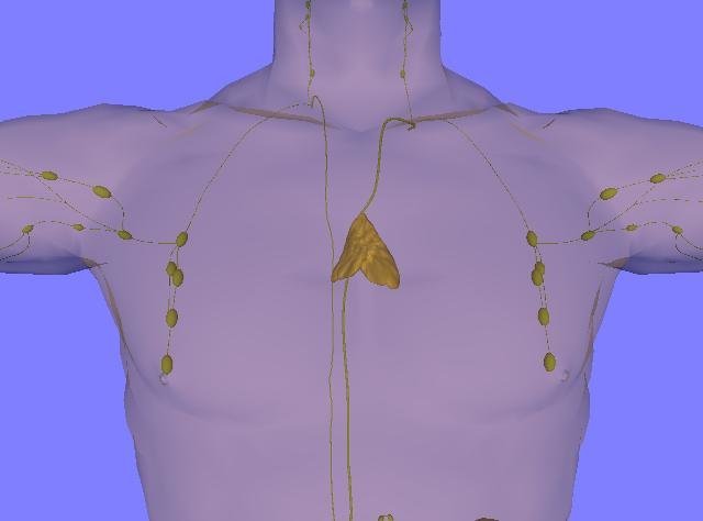
Adult chest. It shows the location and size of the thymus gland in adults. Credit: LearnAnatomy/Wikipedia/CC BY 3.0
Scientists at the La Jolla Institute of Immunology (LJI) are studying a talented type of T cell.
Most T cells function only within the person who created them. T cells fight threats by responding to molecular fragments belonging to pathogens, but only when these molecules are combined with markers originating from their own tissues. Your flu-fighting T cells cannot help your neighbor and vice versa.
“But we all have T cells that don’t follow these rules,” says Dr. Mitchell Cronenberg, LJI professor and president emeritus. “One of these cell types is mucosa-associated invariant T (MAIT) cells.”
Now, Cronenberg and his LJI colleagues have discovered another superpower of MAIT cells. That is, MAIT cells can recognize the same markers, whether derived from humans or mice. Cronenberg called the discovery “remarkable.” “Humans diverged from rats during evolution 60 million years ago,” he says.
This new research published in scientific immunologyreveals the genes and nutrients that give MAIT cells their fighting power. This discovery is an important step toward one day using these cells to treat infectious diseases and improve cancer immunotherapy.
“MAIT cells are the same between individuals, so they can be used more easily for cell therapy. In principle, I could give you my MAIT cells,” Cronenberg says.
This new research also opens the door to utilizing MAIT cells to improve cell therapy. “If we can bring normal T cells closer to MAIT cells, we may be able to make them work faster and more vigorously to fight all types of infections and cancer,” said co-first author of the study. said Gabriel Asqui, a graduate student at LJI at the University of California, San Diego.Cronenberg Institute
What makes MAIT cells special?
Cronenberg was initially interested in the MAIT cell’s unexpected response speed. Typical His T cells require several days to develop within the thymus and adapt to fight new threats only after leaving the thymus and after several days of stimulation from the pathogen. MAIT cells are much faster because they can respond to more general markers of infection rather than looking for very specific tissue type markers. For MAIT cells, a red flag is a red flag no matter who is waving it.
This broad specificity makes MAIT cells similar to the first responder cells of the immune system, such as macrophages and neutrophils, which make up the “innate” immune system. “MAIT cells have ‘innate-like’ characteristics,” Askui says. “They’re like a first line of defense.” In fact, MAIT cells tend to cluster in tissues like the lungs and intestines, where the body is under constant threat from airborne and food-borne pathogens. Masu.
New research shows that MAIT cells not only recognize different markers in one person’s body. Instead, these strange she-T cells can “see” markers that are shared between humans and even between species. Scientists call this type of shared marker “conserved.” There is no reason for markers to change over the years and remain the same between related species.
But just because these MAIT cells look the same across species doesn’t mean they fight pathogens or generate energy in exactly the same way.
Why focus on mouse cells?
Comparing human and mouse MAIT cells is important in guiding future studies that can use mice as a useful animal model to study exactly how these cells fight pathogens.
Kronenberg, Ascui, and their colleagues used single-cell sequencing and other tools to compare differences in gene expression pathways between human and mouse MAIT cells. Researchers discovered that mice have two types of MAIT cells that produce different inflammatory molecules called cytokines. A type of MAIT cell, which scientists call MAIT1, produces large amounts of a cytokine called interferon gamma. Another type of her MAIT cells, called MAIT 17, produces large amounts of a cytokine called interleukin-17.
Recent natural cell biology The Cronenberg Institute study, co-led by LJI Instructor and Immunometabolism Core Director Dr. Tom Riffelmacher, found that after bacterial infection, MAIT1 and MAIT17 cells persist, but become hypercharged or develop stronger defenses. It has been shown that it is possible to have A few months. These cytokines help MAIT cells target a variety of threats. MAIT1 cells target viruses such as influenza, while MAIT17 cells are better at targeting bacteria.
In the new study, the team found that both types of MAIT cells have a greater ability to take up and store fat compared to typical T cells. This finding suggests that MAIT cells are even more dependent on this nutrient for energy. This finding is also consistent with previous research from the Cronenberg Institute that showed that some MAIT cells rely on fat to fight pathogens. The main difference between the two species is that human MAIT cells can produce interferon gamma and IL-17, but separate cell populations clearly cannot.
When rats live like us
Scientists needed to know whether this difference between human and mouse MAIT cells was related to genetic differences or to differences in habitat. Laboratory mice, such as those raised at LJI, are kept in ultra-clean rooms. Their food is autoclaved to kill pathogens, and their water, toys, and cages are kept as sterile as possible.
Cronenberg and Askui were interested in whether mice living in poorly controlled environments showed differences in MAIT cell function. The research team, in collaboration with scientists at the University of California, San Diego, studied MAIT cells from mice kept in so-called “dirty” or worse sterile conditions, similar to a pet store environment. Their research shows that MAIT cells from these mice have even more in common with human MAIT cells, particularly in that MAIT1 cells are more abundant and produce more interferon-gamma than MAIT1 cells from laboratory mice. This suggests that they have something in common.
“Pet stores aren’t dirty in the traditional sense,” Cronenberg said. “But part of the idea is that the ‘dirty’ mice are living in an environment a little closer to the human environment, with more microbial and immune system challenges.”
The researchers also compared MAIT cells in different parts of the body, including the blood, the thymus (where T cells, including MAIT cells, originate), and the lungs and spleen (where MAIT cells camp). They found that the MAIT cells still present in the thymus were very similar in humans and mice (‘dirty’ or not). However, MAIT cells from the lungs and blood are even more different in humans and laboratory mice.
MAIT cells in “dirty” mice were between the two groups, adding further evidence that a more natural environment changes how MAIT cells develop and learn to target disease. Ta.
“Not only genetic differences but also the environment shape the differences between these cell species,” Cronenberg says.
What does this mean for clinical research?
A new study provides scientists with a sort of key to the answer: a list of genetic characteristics that allow MAIT cells to be differentiated according to the species and tissue of their origin. The researchers are now interested in whether they can induce typical T cells to express similar genetic signatures.
“If we can create more ‘innate’ normal cells like MAIT cells, we may be able to improve T-cell therapy for cancer,” Askui says. “That’s one avenue we’re considering.”
Cronenberg is also interested in whether scientists can modify MAIT cells to actually reduce IL-17 levels in the body. IL17 helps fight infections, but some T cells produce IL-17 for the wrong targets, causing harmful inflammation and even autoimmune diseases.
“There are times when IL-17 is the bad guy,” Cronenberg said. “So while we may want to induce more MAIT17 cells and expand that population, we also want to find ways to prevent MAIT17 cells from developing in situations that we may not want. ”
For more information:
Shilpi Chandra et al, Defining mouse and human MAIT cell populations by transcriptome and metabolism, scientific immunology (2023). DOI: 10.1126/sciimmunol.abn8531. www.science.org/doi/10.1126/sciimmunol.abn8531

